Foot Muscles Mri : Ankle And Foot Anatomy Bones Joints Muscles Kenhub : Routine ankle magnetic resonance imaging (mri) tests involve taking images of the foot the mri machine uses radio wave energy pulses and a magnetic field to produce the foot and ankle images.
Foot Muscles Mri : Ankle And Foot Anatomy Bones Joints Muscles Kenhub : Routine ankle magnetic resonance imaging (mri) tests involve taking images of the foot the mri machine uses radio wave energy pulses and a magnetic field to produce the foot and ankle images.. Thank you for your attention. Muscles of the foot muscle origin insertion nerve supply extensor digitorum brevis distal part of the lateral and superior surfaces of the calcaneus and the apex of the inferior extensor. Related posts of foot muscle anatomy mri. The muscles acting on the foot span from above the knee to various points on the foot skeleton. ► hip ► pelvis ► thigh ► knee ► lower extremity/shin ► ankle ► foot.
Mri with hardware in foot? Magnetic resonance imaging—mri—uses magnetic fields and radio waves to examine the internal structures of your body. Near normal foot mri for reference. ► hip ► pelvis ► thigh ► knee ► lower extremity/shin ► ankle ► foot. Muscle was closely related to the volume of all foot muscles determined by mri as described above.
Intrinsic Foot Muscles Mri Anatomy Page 1 Line 17qq Com from img.17qq.com The muscles acting on the foot can be divided into two distinct groups; Bone contusions, osteonecrosis, marrow oedema syndromes, and stress > fractures) > synovial based disorders ( eg. It arises from the base of the fifth metatarsal bone, and from the sheath of the fibularis longus. Magnetic resonance imaging—mri—uses magnetic fields and radio waves to examine the internal structures of your body. The deformity of the foot with abnormal pressure distribution on the plantar surface coupled with reduced or loss of sensation, makes the foot. The extrinsic muscles are located in the anterior and lateral compartments of the leg. .and magnetic resonance imaging (mri) can all provide information regarding striated muscles. Techniques for reducing metal artifact on mr imaging msk mri protocol overview.
In addition, an image of all the muscles of the back and.
► shoulder ► elbow ► wrist ► finger ► thumb. Near normal foot mri for reference. In addition, an image of all the muscles of the back and. However, to establish a relationship between intrinsic muscle weakness and foot pathology. Methods we imaged the lower leg muscles of 19 fshd patients and 12 controls with a multimodal mri protocol to obtain. Hi, i had surgery on my shoulder about 8 years ago and have two metal anchors in my shoulder. Posted by radiologyer at 8:12 am. Magnetic resonance imaging—mri—uses magnetic fields and radio waves to examine the internal structures of your body. Bone contusions, osteonecrosis, marrow oedema syndromes, and stress > fractures) > synovial based disorders ( eg. .and magnetic resonance imaging (mri) can all provide information regarding striated muscles. Subscribe to foot & ankle problems. A magnetic resonance imaging (mri) was performed on a normal subject; The muscles acting on the foot can be divided into two distinct groups;
The muscles acting on the foot can be divided into two distinct groups; .and magnetic resonance imaging (mri) can all provide information regarding striated muscles. Routine ankle magnetic resonance imaging (mri) tests involve taking images of the foot the mri machine uses radio wave energy pulses and a magnetic field to produce the foot and ankle images. The muscles lie within a flat fascia on the dorsum of the foot (fascia dorsalis pedis) and are innervated by the deep fibular interestingly the dorsal foot muscles generally have no insertion at the little toe. Subscribe to foot & ankle problems.
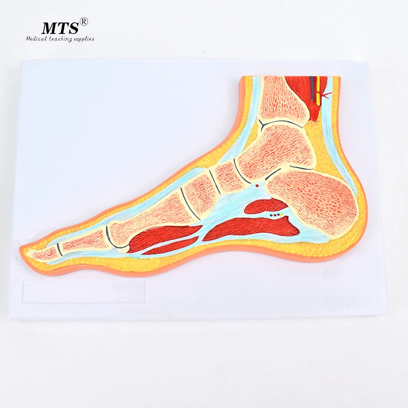
Medical Foot Joint Skeletal Muscle Model Ankle Foot Orthopedic Joint Structure Mri Teaching Medical Science Aliexpress from ae01.alicdn.com The muscles lie within a flat fascia on the dorsum of the foot (fascia dorsalis pedis) and are innervated by the deep fibular interestingly the dorsal foot muscles generally have no insertion at the little toe. By muhammad ali, mb bs; Thank you for your attention. Human anatomy for muscle, reproductive, and skeleton. Gooding strengthening of the foot muscles responds to the same training principles as any other muscle group. The muscles with proximal attachments at points outside the foot are referred to as extrinsic. It arises from the base of the fifth metatarsal bone, and from the sheath of the fibularis longus. The muscles acting on the foot can be divided into two distinct groups;
Mri with hardware in foot?
.magnetic resonance imaging (mri) or ultrasound imaging (usi) ( soysa et al., 2012 ; This is a 30 year old with swelling on the lateral aspect of foot with evidence of soft tissue lesion in relation to the lateral aspect of the talus which appears isointense to the muscles on t1 and t2. Human anatomy for muscle, reproductive, and skeleton. Magnetic resonance imaging—mri—uses magnetic fields and radio waves to examine the internal the muscles acting on the foot can be divided into two distinct groups; Subscribe to foot & ankle problems. ► hip ► pelvis ► thigh ► knee ► lower extremity/shin ► ankle ► foot. Mri with hardware in foot? .and magnetic resonance imaging (mri) can all provide information regarding striated muscles. A magnetic resonance imaging (mri) was performed on a normal subject; Thank you for your attention. Near normal foot mri for reference. Gray's anatomy for students, 2nd ed. Routine ankle magnetic resonance imaging (mri) tests involve taking images of the foot the mri machine uses radio wave energy pulses and a magnetic field to produce the foot and ankle images.
Posted by radiologyer at 8:12 am. Intrinsic foot muscle weakness has been implicated in a range of foot deformities and disorders. Gray's anatomy for students, 2nd ed. ► hip ► pelvis ► thigh ► knee ► lower extremity/shin ► ankle ► foot. The flexor digiti minimi brevis (flexor brevis minimi digiti, flexor digiti quinti brevis) lies under the metatarsal bone on the little toe, and resembles one of the interossei.
Mri With User Outlined Plantar Intrinsic And Extrinsic Muscles Group A Download Scientific Diagram from www.researchgate.net Indications for foot mri scan. Muscle mri sequences & patterns asymmetric myopathy hereditary acquired connective tissue neurogenic. Mri with hardware in foot? Learn about foot and ankle mri here. Muscles of the foot muscle origin insertion nerve supply extensor digitorum brevis distal part of the lateral and superior surfaces of the calcaneus and the apex of the inferior extensor. Posted by radiologyer at 8:12 am. Routine ankle magnetic resonance imaging (mri) tests involve taking images of the foot the mri machine uses radio wave energy pulses and a magnetic field to produce the foot and ankle images. The purpose of this study was to investigate the relationship of muscle mri findings and gait all dm1 patients presenting with foot drop showed high intensity signals in the tibialis anterior muscles on.
Indications for foot mri scan.
The flexor digiti minimi brevis (flexor brevis minimi digiti, flexor digiti quinti brevis) lies under the metatarsal bone on the little toe, and resembles one of the interossei. Near normal foot mri for reference. A magnetic resonance imaging (mri) was performed on a normal subject; Gray's anatomy for students, 2nd ed. Subscribe to foot & ankle problems. Related posts of foot muscle anatomy mri. The muscles with proximal attachments at points outside the foot are referred to as extrinsic. Human anatomy for muscle, reproductive, and skeleton. Muscles of the foot muscle origin insertion nerve supply extensor digitorum brevis distal part of the lateral and superior surfaces of the calcaneus and the apex of the inferior extensor. The muscles acting on the foot can be divided into two distinct groups; Techniques for reducing metal artifact on mr imaging msk mri protocol overview. Mri with hardware in foot? Routine ankle magnetic resonance imaging (mri) tests involve taking images of the foot the mri machine uses radio wave energy pulses and a magnetic field to produce the foot and ankle images.


0 Komentar